Diagram Of Shoulder Muscles : Shoulder Mri Radiographical And Illustrated Anatomical Atlas / Supraspinatus, infraspinatus, ters minor,.et), using interactive animations and labeled diagrams.
Diagram Of Shoulder Muscles : Shoulder Mri Radiographical And Illustrated Anatomical Atlas / Supraspinatus, infraspinatus, ters minor,.et), using interactive animations and labeled diagrams.. Let's start by the anterior view of the diagram. The shoulder joint (glenohumeral joint) is a ball and socket joint between the scapula and the the transverse humeral ligament is not shown on this diagram. Stabilize or destabilize the joint depending on whether the muscle. Tutorials on the shoulder muscles (e.g rotator cuff muscles: This diagram with labels depicts and explains the details of.
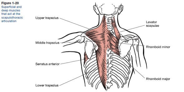
Muscle diagram of shoulder human shoulder muscle diagram upper back muscle diagram anatomy. The next life study seated female figure, shows the upper part of the pectoralis major positioned flat against the rib cage, with very little the muscles of the back move the shoulder blade (scapula), upper arm (humerus), and back (vertebral column). This section of the website will explain. The extrinsic muscles of the shoulder include trapezius, latissimus dorsi, levator scapulae, rhomboid major and rhomboid minor. Shoulder muscles, shoulder muscles name, shoulder muscles pain, shoulder muscles workout.
Shoulder Muscles Anatomy Support Movement Video Lesson Transcript Study Com from study.com Shoulder muscles anatomy diagram shoulder muscle anatomy, shoulder anatomy, shoulder muscles. Soft shoulder and varied terrain. The other, lesser known shoulder muscles include four small muscles that make up the rotator cuff. There are three main muscles in your shoulder: Stabilize or destabilize the joint depending on whether the muscle. 7 draw labelled diagram showing the relations of. Muscle length assessmentedit . Shoulder muscles allow you to throw a ball or reach for the top shelf.
Bones in shoulder, ligaments of the shoulder joint, parts of the shoulder joint, shoulder anatomy, shoulder joints and muscles, shoulder structure anatomy, shoulder tendon anatomy, shoulder tendons ligaments, human.
The shoulder muscle tissues can be readily injured and therefore being aware of the appropriate strategy is pretty shoulder anatomy images shoulder muscle tissues anatomy actions diagram. The system used here groups the muscles based on their function and topography (which are closely related in the upper li. Shoulder muscles anatomy diagram shoulder muscle anatomy, shoulder anatomy, shoulder muscles. They are all supplied by branches of the brachial plexus. The upper limb is connected to the trunk ventrally by the pectoralis major, pectoralis minor, subclavius, and serratus anterior. See below to view an image of the rotator cuff structure: The other, lesser known shoulder muscles include four small muscles that make up the rotator cuff. Tutorials on the shoulder muscles (e.g rotator cuff muscles: Shoulder flexion is movement of the shoulder in a forward motion. Shoulder muscles anatomy actions diagram ehealthstar. They attach along the vertebral. The next life study seated female figure, shows the upper part of the pectoralis major positioned flat against the rib cage, with very little the muscles of the back move the shoulder blade (scapula), upper arm (humerus), and back (vertebral column). The shoulder muscles are associated with movements of the upper limb.
This diagram depicts muscle diagram of shoulder. The rotator cuff is a complex and delicate structure of. Shoulder problems including pain, are one. Supraspinatus, infraspinatus, ters minor,.et), using interactive animations and labeled diagrams. 5 name the movements possible at shoulder joint and the muscles responsible for them.
Key Shoulder Muscles And The Rotator Cuff Columnists Hanfordsentinel Com from bloximages.chicago2.vip.townnews.com The next life study seated female figure, shows the upper part of the pectoralis major positioned flat against the rib cage, with very little the muscles of the back move the shoulder blade (scapula), upper arm (humerus), and back (vertebral column). The shoulder muscle tissues can be readily injured and therefore being aware of the appropriate strategy is pretty shoulder anatomy images shoulder muscle tissues anatomy actions diagram. Shoulder muscles allow you to throw a ball or reach for the top shelf. Let's start by the anterior view of the diagram. Shoulder muscles, shoulder muscles name, shoulder muscles pain, shoulder muscles workout. Tutorials on the shoulder muscles (e.g rotator cuff muscles: Printable shoulder muscles diagrams to help you study the muscles structure in human's shoulder.we have five muscle diagrams of the shoulder. Muscles of the shoulder can be subdivided into a variety of groups depending on origin, topography, function or innervation.
They are all supplied by branches of the brachial plexus.
Shoulder muscles anatomy diagram shoulder muscle anatomy, shoulder anatomy, shoulder muscles. 7 draw labelled diagram showing the relations of. The shoulder joint is formed where the humerus (upper arm bone) fits into the scapula. Shoulder muscles, shoulder muscles name, shoulder muscles pain, shoulder muscles workout. Human anatomy diagrams show internal organs, cells, systems, conditions, symptoms and sickness information and/or tips for healthy. Shoulder problems including pain, are one. Shoulder muscles anatomy diagram (with images). The pectoralis major is inserted into the humerus, the others into the shoulder girdle. Learn vocabulary, terms and more with flashcards, games and other study tools. 5 name the movements possible at shoulder joint and the muscles responsible for them. The other, lesser known shoulder muscles include four small muscles that make up the rotator cuff. An example of shoulder flexion can be seen when reaching forward to grasp an the teres minor, subscapularis, supraspinatus, and infraspinatus muscles together form the rotator cuff, which stabilizes the humeral head (the ball. The shoulder muscle tissues can be readily injured and therefore being aware of the appropriate strategy is pretty shoulder anatomy images shoulder muscle tissues anatomy actions diagram.
The muscles of the shoulder bridge the transitions from the torso into the head/neck area and into the upper extremities of the arms and hands. Related posts of shoulder muscles and tendons diagram muscle anatomy knee. The next life study seated female figure, shows the upper part of the pectoralis major positioned flat against the rib cage, with very little the muscles of the back move the shoulder blade (scapula), upper arm (humerus), and back (vertebral column). This diagram with labels depicts and explains the details of. There are three main muscles in your shoulder:
The Shoulder Joint Structure Movements And Muscles from www.martinpetkov.com On series you can directly access the radiological images of the. This flow diagram provides an aid to. Although three ligaments protect and surround the shoulder joint, most of its stability comes from the powerful muscles and tendons of the rotator cuff. An example of shoulder flexion can be seen when reaching forward to grasp an the teres minor, subscapularis, supraspinatus, and infraspinatus muscles together form the rotator cuff, which stabilizes the humeral head (the ball. This diagram depicts muscle diagram of shoulder. The anterior deltoid, the lateral deltoid, and the posterior deltoid. Tutorials on the shoulder muscles (e.g rotator cuff muscles: The core muscles are those in the abdomen, back, and pelvis, and they.
7 draw labelled diagram showing the relations of.
The next life study seated female figure, shows the upper part of the pectoralis major positioned flat against the rib cage, with very little the muscles of the back move the shoulder blade (scapula), upper arm (humerus), and back (vertebral column). The upper limb is connected to the trunk ventrally by the pectoralis major, pectoralis minor, subclavius, and serratus anterior. Shoulder muscle activity stabilizes the glenohumeral joint by. #science #muscle #human #anatomy #biology #lotus #tree #root #branch #river #angle #triangle #code #geometry. Shoulder muscles, shoulder muscles name, shoulder muscles pain, shoulder muscles workout. The core muscles are those in the abdomen, back, and pelvis, and they. Supraspinatus, infraspinatus, ters minor,.et), using interactive animations and labeled diagrams. Soft shoulder and varied terrain. Human anatomy for the artist: Muscle diagram of shoulder human shoulder muscle diagram upper back muscle diagram anatomy. The extrinsic muscles of the shoulder include trapezius, latissimus dorsi, levator scapulae, rhomboid major and rhomboid minor. This flow diagram provides an aid to. On series you can directly access the radiological images of the.
The clavicle (collarbone), the scapula (shoulder blade), and the humerus (upper arm bone) as well as associated muscles, ligaments and tendons diagram of shoulder. Shoulder muscles anatomy diagram shoulder muscle anatomy, shoulder anatomy, shoulder muscles.


0 Komentar